


A plant ovule, the precusor to a plant seed.
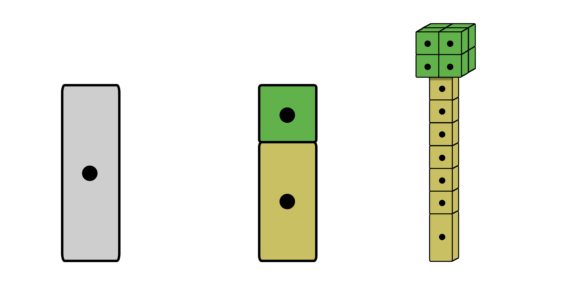
First stages of a zygote dividing to eventually form a mature plant embryo (not shown).
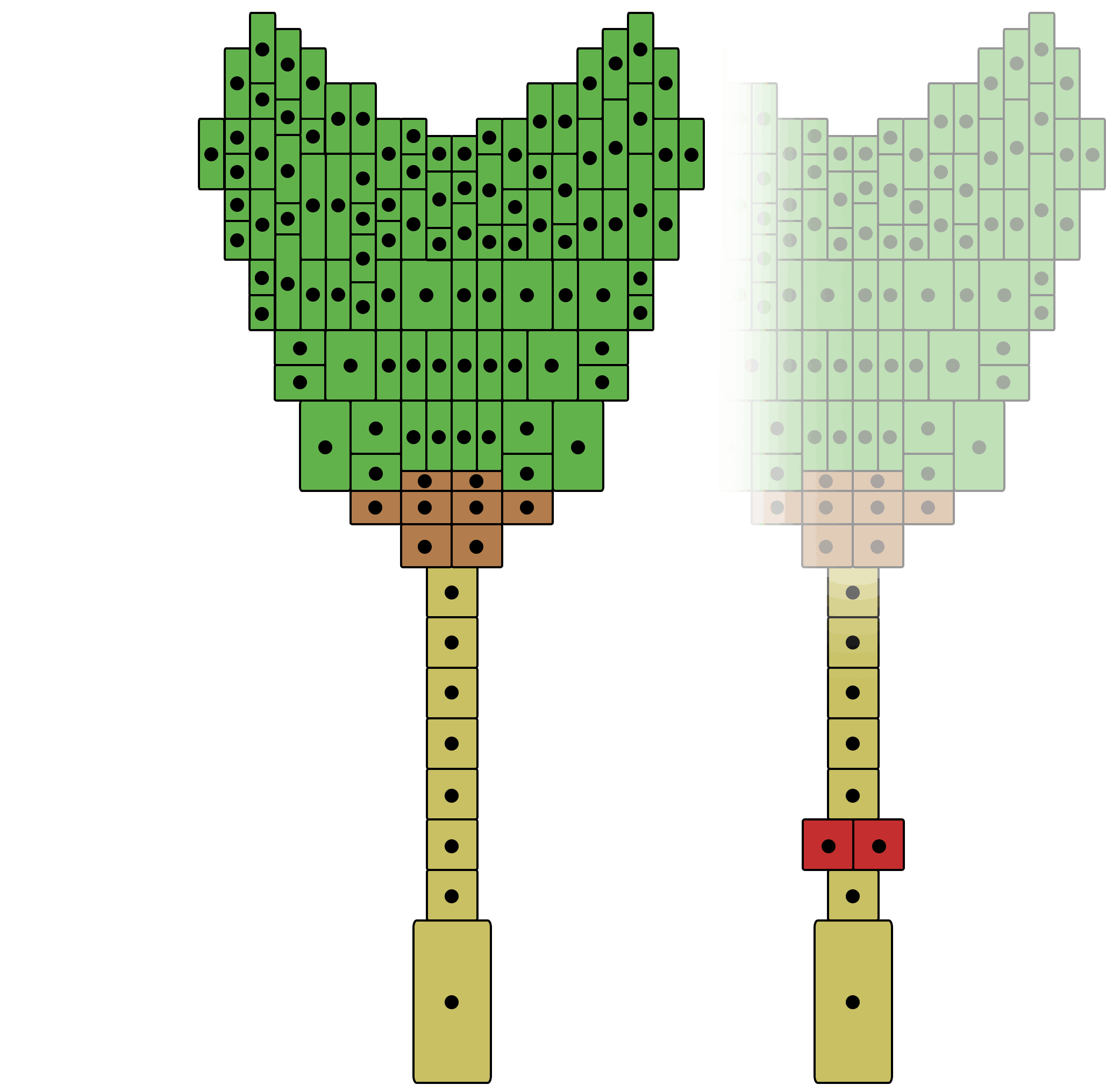
A heart stage embryo in which a wrong division occurred in the suspensor organ.
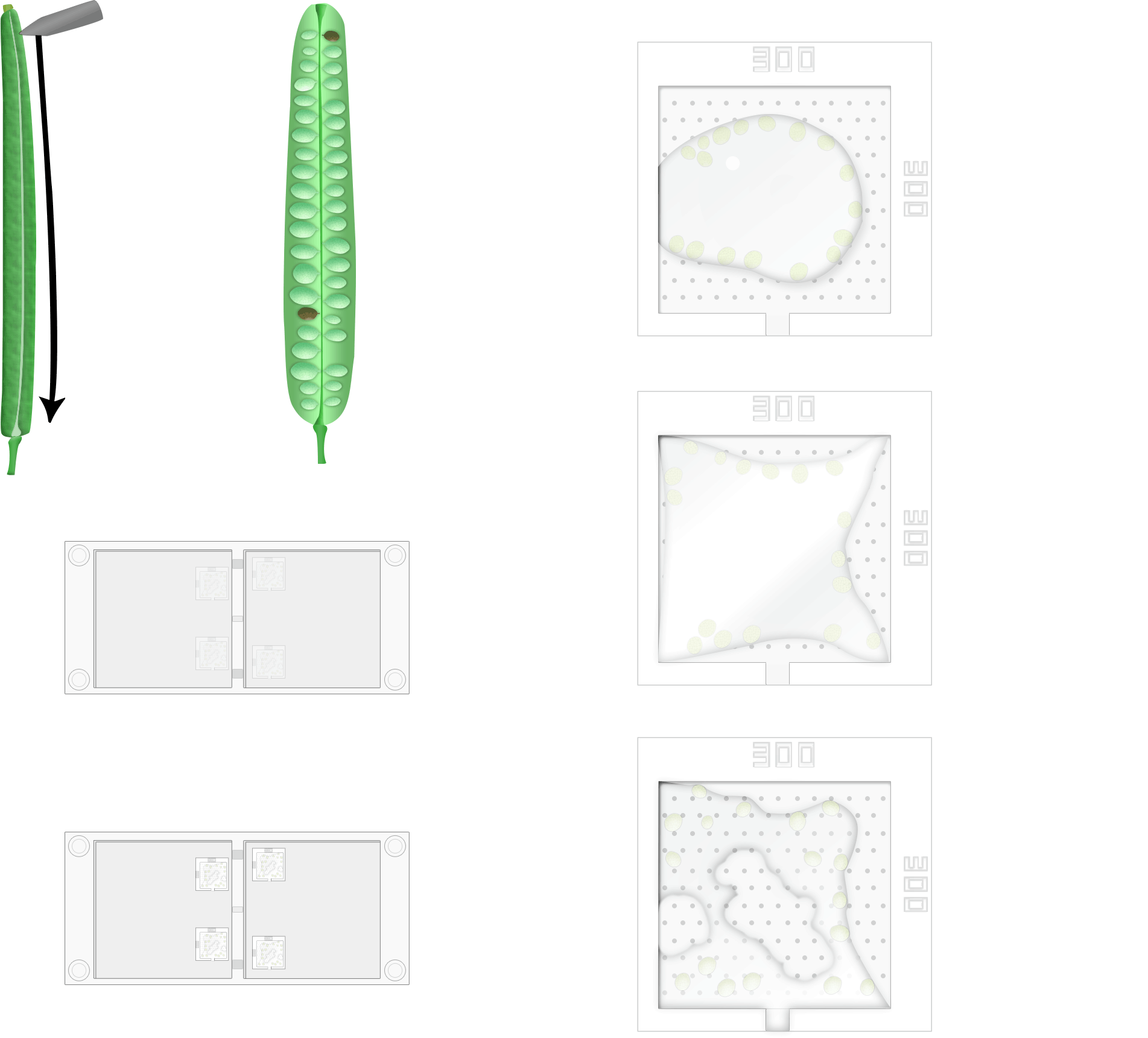
A highly precise procedure to isolate living ovules from Arabidopsis Thaliana siliques. Isolated ovules are secured in liquid medium inside a polydimethylsiloxane micropillar device, this device is used to mount ovules in place. Mounted ovules are then live imaged to capture suspensor cell divisions by using fluorescence confocal microscopy.
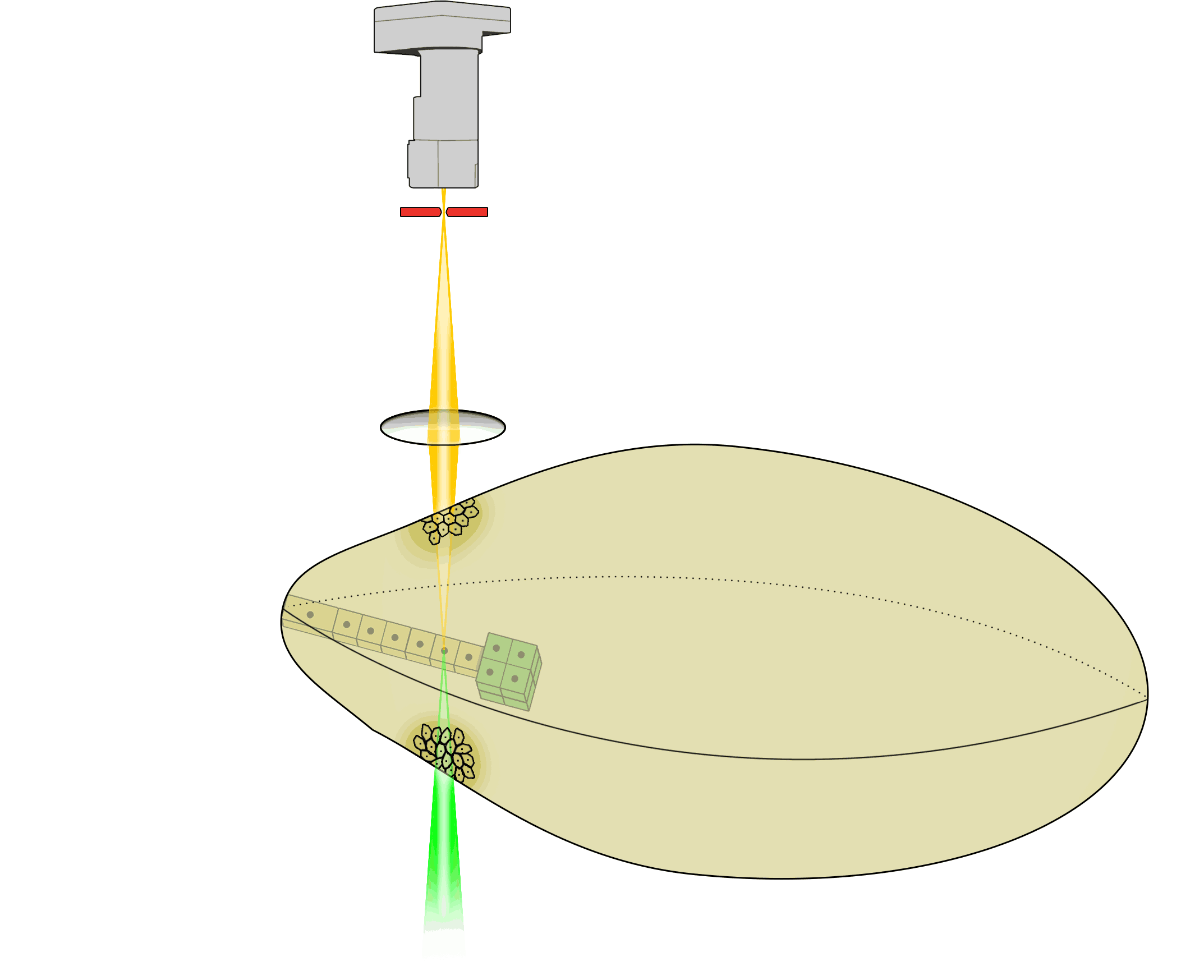
Basic explanation of fluorescence confocal microscopy: the suspensor organ inside a living ovule is excited by focusing green laser light onto it. Excited yellow fluorescent protein inside cells of the suspensor then re-emit yellow light which is focused through a pinhole by a convex lens and eventually detected by a highly sensitive hybrid detector.